Thursday, May 25, 2023

The Virtual Mummy: Unwrapping a Mummy by Mouse Click

[First posted in AWOL 29 November 2014, updated 25 May 2023]

The Virtual Mummy: Unwrapping a Mummy by Mouse Click

Head of the VIrtual Mummy
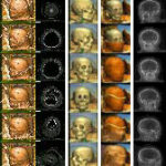
The computer has entered our everyday life and did not stop before the field of mummy research. It helps researchers not only in the non-destructive examination of mummies, but even allows the creation of a “virtual mummy”, which visitors of the exhibition The Secret of the Mummies – Eternal Life at the Nile, but also you (with limited functionality) can unwrap on the screen. The object is a 2300-year-old mummy of a female, aged about 30 years.

Naturally, the unwrapping by computer is no longer the mysterious and thrilling experience that a real unwrapping used to be. But according to modern opinion, such a procedure, which disregards the dignity of the deceased, would no longer be acceptable anyway. This problem does not arise with the virtual mummy.

The procedure originated from medical research. The initial aim was to provide a highly accurate visualization of the interior body from tomographic images to improve medical diagnostics, surgical planning, and education of medical students.

For more than 12 years, a group of researchers headed by Professor Karl Heinz Höhne, PhD, at the Institute of Mathematics and Computer Science in Medicine (IMDM) has done research in the field of anatomical 3D reconstruction of the living human body. Procedures have improved over the years, and thus, it is nowadays possible to produce computer-based body models (“virtual bodies”), which can be examined in the way an anatomist or surgeon would do it. With the program named VOXEL-MAN, operations can be simulated and planned in advance, and 3D anatomical atlases can be produced. In these fields, Höhne’s group did pioneering work, like for example in 1987, when the first brain of a living human being was reconstructed. Meanwhile, the focus in on the development of medical training simulators.

The computer, of course, does not mind whether the reconstructed object is a patient or a mummy. It was at hand to examine the latter using the same procedure. The image quality is usually even better, as high doses of radiation can be applied without risk, and the mummy patiently holds still.

The application for mummies was developed in cooperation with Renate Germer, PhD, from the Dept. of Egyptology, University of Hamburg. Work began in 1989 and was first presented in 1991 at the exhibition Mumie + Computer at the Kestner Museum, Hanover. The web pages were published in 1997.


No comments:

Post a Comment